Cardiologie
De dienst cardiologie behandelt aandoeningen van hart- en bloedvaten (cardiovasculaire aandoeningen of hart- en vaatziekten).
Praktisch
Maak een afspraak in het UZA
UZA Cardiologie
Niet beschikbaar
Maak een afspraak in je buurt
Stafleden van de dienst cardiologie doen raadplegingen in het UZA, maar ook op een aantal andere locaties in de Antwerpse regio. Zo vind je steeds een UZA-cardioloog in je buurt.
Praktijk Epione, Buizegem Edegem
Praktijk Willebroek
Diagnosecentrum Voorkempen | Zoersel
Nottebohm Medisch Centrum Antwerpen | Prof. Van Craenenbroeck
Nottebohm Medisch Centrum Antwerpen | Dr. De Roeck
Nottebohm Sport Medisch Centrum Brecht
Cardiologie Ziekenhuis Geel
Binnenkort naar het UZA?
Heb je binnenkort een afspraak bij het UZA of kom je iemand bezoeken? Beantwoord enkele vragen en krijg handige tips voor je UZA-bezoek, van thuisvoorbereiding tot navigatie in het ziekenhuis.
Wat is cardiologie?
De dienst cardiologie behandelt aandoeningen van hart- en bloedvaten (cardiovasculaire aandoeningen of hart- en vaatziekten).
De dienst wil zich zowel nationaal als internationaal onderscheiden door een voortdurende bevordering van de kwaliteit van individuele patiëntenzorg, wetenschappelijk onderzoek en academisch onderwijs. De multidisciplinaire en innovatieve aanpak van het cardiologisch team is de garantie om onze taak als topreferentiecentrum in cardiovasculaire aandoeningen waar te maken.
Hiervoor kan je bij ons terecht
Subspecialismen

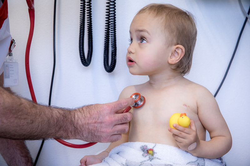 De dienst kindercardiologie van het UZA centraliseert de zorg voor kinderen met aangeboren of verworven hartafwijkingen.
Hartkatheterisatielabo
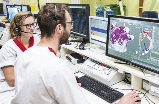
Hartkatheterisatielabo
Hartpatiënten kunnen hier terecht voor onderzoek en behandeling van angina pectoris of hartaanval, hartkamer- en klepaandoeningen, of hartritmestoornissen.
Poliklinieken

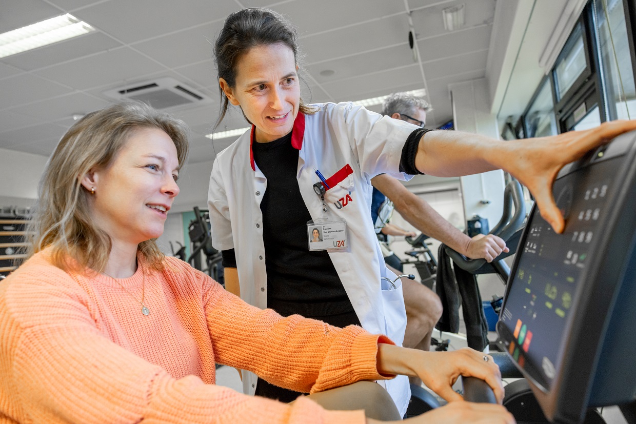 Met de Vrouw en hart-raadpleging wil het UZA hartkwalen bij vrouwen vroeger opsporen en behandelen.
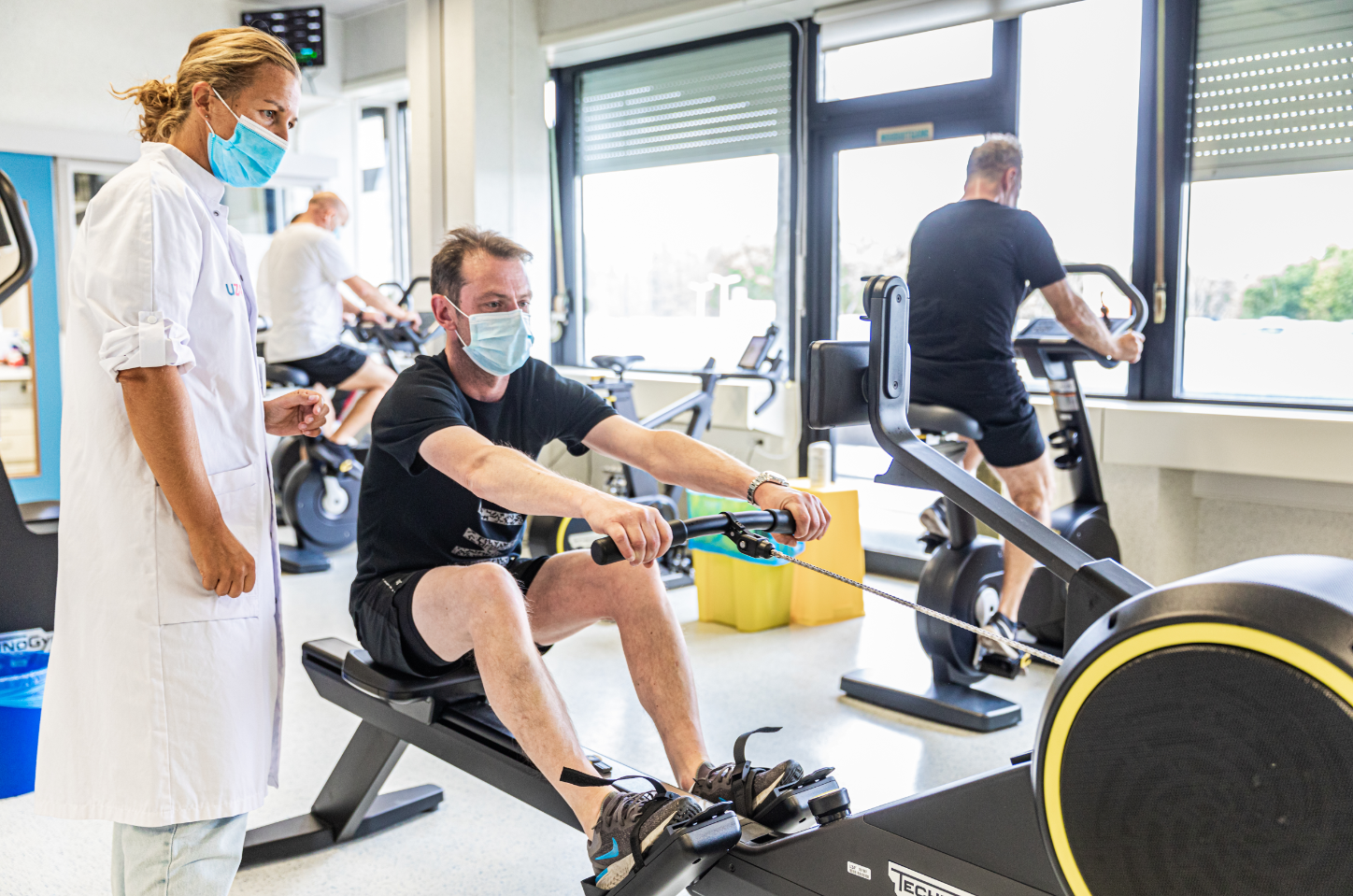 Onderzoek heeft aangetoond dat cardiale revalidatie een positief effect heeft op de levenskwaliteit van hartpatiënten.
Zorgaanbod
Aandoeningen
Onderzoeken en behandelingen
Psychologische zorg op de dienst cardiologie
Patiënten die in een behandelplan zitten van cardiale revalidatie kunnen rekenen op gespecialiseerde psychologische zorg, gericht op diverse hulpvragen die kunnen ontstaan tijdens de cardiale revalidatie, zoals verwerkingsproblemen, aanpassingsmoeilijkheden bij het leven met een medisch apparaat, trauma, verlieservaringen (rouw en verdriet), en arbeidsre-integratie. Psychologen bieden hulp aan zowel gehospitaliseerde als ambulante patiënten. Dit omvat individuele sessies en infosessies met aanvullende informatie en ondersteuning.
Onze zorgverleners staan voor je klaar
Hieronder vind je een overzicht van de zorgverleners die deel uitmaken van ons team en hun specifieke rol in de behandeling en ondersteuning van onze patiënten.

Artsen


Consulenten en geassocieerde artsen


Coördinatoren

Verpleegkundigen


Paramedici


Psychologen

Patiëntenbegeleiding


Word jij onze collega?
Arts, jobstudent, verpleegkundige, labotechnicus, onthaalmedewerker, schoonmaakmedewerker, … Bij het UZA delen we allemaal momenten die ertoe doen. Elke dag anders, maar altijd samen.
Dit is jouw moment. Vind hier je job met impact.
Klinische studies
Elk jaar nemen er in het UZA heel wat patiënten deel aan klinische studies. Dit gebeurt op vrijwillige basis. Indien er op de dienst waar je bent opgenomen een specifiek onderzoek loopt, zal je eventueel gevraagd worden om hieraan deel te nemen. Je hebt de volledige vrijheid om te beslissen of je hier al dan niet op in wenst te gaan. Indien je deelneemt of in de toekomst deelneemt aan klinische studies, kan je met je vragen terecht bij je behandelend arts.


 bij cardiologie")
